
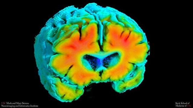
New research from a major neuroimaging program suggests that the brain’s ability to regulate blood flow and deliver oxygen may be tied to features commonly seen along the Alzheimer’s disease pathway. Using resting-state, noninvasive tools that are already common in hospitals and research centers, the study links healthier vascular regulation to fewer amyloid plaques and larger hippocampal volume—markers that generally track with better cognitive health. Published in a peer‑reviewed study in Alzheimer’s and Dementia in 2026, the work comes from the Mark and Mary Stevens Neuroimaging and Informatics Institute at the Keck School of Medicine of USC and underscores how vascular function could complement established neurodegeneration measures. The authors emphasize that the analysis captures a single point in time and is not proof of causation.
Vascular signals tied to hallmark Alzheimer’s brain changes
The findings land in the context of long‑standing evidence that the vascular system and the brain are tightly intertwined: arteries, veins and capillaries supply the oxygen and nutrients that support neuronal function, and vascular disease is already recognized as an important contributor to stroke and cognitive decline. Against that backdrop, the USC team set out to quantify how moment‑to‑moment regulation of blood flow and oxygenation relates to structural and molecular markers of Alzheimer’s‑related change.
Researchers assessed two signals while participants rested quietly: blood‑flow velocity in major brain arteries and oxygenation in superficial cortical tissue. Statistical models summarized how well each person’s cerebrovascular system adjusted to natural fluctuations in blood pressure and carbon dioxide. Higher values—indicating vessel behavior closer to cognitively healthy aging—tracked with brain features associated with lower Alzheimer’s risk.
“Amyloid and tau are often considered the primary players in Alzheimer’s disease, but blood flow and oxygen delivery are also critical,” said Amaryllis A. Tsiknia, lead author of the study and USC PhD candidate. “Our results show that when the brain’s vascular system functions more like it does in healthy aging, we also see brain features that are linked to better cognitive health.”
“These vascular measures are capturing something meaningful about brain health,” said Meredith N. Braskie, PhD, senior author of the study and assistant professor of neurology at the Keck School of Medicine. “They appear to align with what we see on MRI and PET scans that are commonly used to study Alzheimer’s disease, providing important information about how vascular health and standard brain measures of Alzheimer’s disease risk may be related.”
What the study measured, in brief
The study focused on two noninvasive technologies that are already familiar in many neurology and critical‑care units, but here were repurposed to probe dementia‑related risk.
| Measure | Modality | What it captures | Patient participation | Radiation/contrast |
|---|---|---|---|---|
| Cerebral blood‑flow velocity | Transcranial Doppler ultrasound (TCD) | Beat‑to‑beat flow in large intracranial arteries; dynamic vessel reactivity | Passive rest; probe placement on scalp/temple | None |
| Cortical tissue oxygenation | Near‑infrared spectroscopy (NIRS) | Relative oxy‑/deoxy‑hemoglobin near cortical surface; oxygen delivery/utilization | Passive rest; sensors on scalp | None |
Key findings at a glance
- Higher (healthier) vascular regulation indicators were linked to:
- Lower amyloid plaque burden on established imaging biomarkers
- Larger hippocampal volume, a structure essential for memory
- Participants with mild cognitive impairment or dementia showed poorer vascular indicators than cognitively normal peers.
- The analysis was cross‑sectional; it identifies associations but does not establish cause and effect, and the authors caution that reverse or bidirectional relationships are possible.
Health‑system implications: scalability without injections or scanners
For health‑system leaders facing rising dementia prevalence and limited advanced imaging capacity, the technology mix is notable. The tools used in this research are portable, do not require radiation or contrast, and place minimal demands on patients. That profile makes them attractive for large observational studies, pragmatic trials and community‑based screening initiatives in which full MRI or PET work‑ups for every participant are neither feasible nor affordable.
| Modality | Primary use in brain health | Portability | Patient burden | Relative cost | Throughput potential |
|---|---|---|---|---|---|
| TCD (ultrasound) | Vessel reactivity and flow dynamics | High (cart/bedside) | Low | Low | High |
| NIRS | Cerebral oxygenation patterns | High (wearable/cap) | Low | Low | High |
| MRI | Structure, perfusion, diffusion | Low (fixed suite) | Moderate (scanner time, noise) | High | Low–Moderate |
| PET (including amyloid) | Molecular pathology imaging | Low (fixed suite; radiotracer logistics) | Moderate (injection, radiation) | High | Low |
“These findings add to growing evidence that Alzheimer’s involves meaningful vascular contributions in addition to classic neurodegenerative changes,” said Arthur W. Toga, PhD, director of the Stevens INI. “Understanding how blood flow and oxygen regulation interact with amyloid and brain structure opens new doors for early detection and potentially prevention.”
Regulatory and reimbursement landscape (United States)
Because the technologies examined in the study are already present in many hospitals, any future shift from research to routine cognitive‑risk assessment would depend less on device approval and more on how payers and professional societies choose to recognize vascular metrics in formal care pathways. In the United States, the U.S. Food and Drug Administration regulates these tools under the existing medical device framework, while coverage decisions are made by public and private insurers and shaped by specialty‑society guidelines.
| Technology | Regulatory status | Billing/coverage context | Relevance to cognitive disorders |
|---|---|---|---|
| Transcranial Doppler (TCD) | FDA Class II devices cleared via 510(k) for diagnostic ultrasound under the broader medical‑device framework set out in the U.S. Food and Drug Administration’s medical device regulations | Established CPT codes for cerebrovascular evaluation; coverage varies by indication and payer | Routine use in stroke, vasospasm, and sickle cell; emerging research in cognitive aging |
| Near‑infrared spectroscopy (cerebral oximetry) | FDA Class II tissue oximeters cleared via 510(k) | Common in perioperative monitoring; coverage for dementia risk assessment not established | Potential adjunct for large research cohorts and community screening studies |
| MRI | FDA‑cleared systems and sequences | Widely covered for neurological evaluation when medically necessary | Standard for structural assessment (e.g., hippocampal atrophy) |
| PET (including amyloid tracers) | FDA‑approved radiopharmaceuticals | Coverage subject to payer policies and clinical criteria; access varies by region | Key role in characterizing amyloid pathology in selected patients and studies |
Public‑health considerations
Policy makers and health‑system planners are increasingly focused on earlier, more equitable detection of cognitive decline. If validated longitudinally, vascular metrics of the type used in this study could become one more instrument in that toolkit, particularly in settings where advanced imaging capacity is scarce.
- Population impact
- Portable vascular measures could help pre‑screen large cohorts, easing bottlenecks for costly imaging slots.
- Lower patient burden may expand participation among older adults with mobility limits or scanner intolerance.
- Community clinics and rural health systems could integrate bedside tools into aging and cognitive‑health programs.
- System capacity
- Potential to standardize acquisition protocols for multi‑site studies and registries.
- Training pathways for technicians and sonographers would be needed to ensure reproducibility.
- Equity and access
- Mobile and home‑based assessments may reduce travel barriers for underserved populations.
- Clear communication is essential to avoid overinterpreting screening signals as diagnosis.
How this fits with established risk frameworks
The work echoes a growing consensus that Alzheimer’s risk sits at the intersection of neurodegeneration, vascular health and lifestyle factors, rather than being driven by a single pathway alone. That framing aligns with existing dementia‑risk guidelines that already emphasize cardiovascular prevention as brain health policy.
- The associations align with the concept that vascular health intersects with neurodegeneration rather than sitting apart from it.
- Well‑known, modifiable risk factors that also influence cerebrovascular health include:
- Hypertension and other cardiovascular disease
- Diabetes and metabolic syndrome
- Tobacco exposure
- Physical inactivity
- Untreated hearing loss
- Any eventual clinical use would need to complement, not replace, established diagnostic work‑ups and biomarker frameworks, including neuropsychological testing, imaging and, where appropriate, cerebrospinal fluid or plasma biomarkers.
What still needs to be proven
For hospital executives, payers and regulators, the central question is whether these vascular signals actually change clinical decision‑making or outcomes. The authors are explicit that the present work is an early‑stage association study; the next phase will need to demonstrate predictive value and clarify how such measures should be embedded in care pathways or population‑health programs.
- Timelines
- 2026: Cross‑sectional analysis published in Alzheimer’s and Dementia (DOI: 10.1002/alz.71146).
- 2026: Longitudinal studies initiated to test whether vascular measures predict future decline or track response to interventions.
- Health outcomes
- Prospective validation that vascular indicators forecast incident cognitive impairment.
- Definition of thresholds, quality controls, and reference ranges across diverse populations.
- Policy measures
- Guidance on when and how to deploy vascular metrics within dementia risk assessments.
- Coverage determinations tied to demonstrated clinical utility and standardized reporting.
“If we can track these signals over time, we may be able to identify people at higher risk earlier and test whether improving vascular health can slow or reduce Alzheimer’s‑related brain changes,” Tsiknia said. For now, the study adds weight to a policy message that is already familiar in cardiovascular medicine but increasingly central to neurology as well: protecting the blood vessels that feed the brain may be one of the most practical levers societies have to influence dementia risk at scale.
