
Transferrin measurement is moving beyond absorbance-only workflows
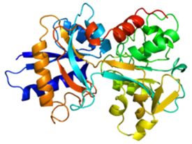
Transferrin is a liver-synthesized glycoprotein that transports iron to cells. Each molecule binds up to two iron atoms and recycles rapidly through the transferrin-receptor pathway, enabling repeated iron delivery cycles during its roughly eight‑day half‑life in humans. This biology underpins routine use of transferrin and transferrin saturation in evaluating iron metabolism across oncology, chronic inflammatory disease, and nutrition programs, and it is increasingly relevant as health systems look for more sensitive tools to detect iron imbalance earlier and in smaller sample volumes.
What a recent instrument comparison found
A bench comparison shared by the manufacturer indicates that a dual‑mode spectrofluorometer detected transferrin at substantially lower concentrations than a low‑volume benchtop spectrophotometer using UV absorbance alone. While this is a single‑manufacturer study and not a head‑to‑head regulatory trial, the performance gap is material for laboratories working with scarce specimens or low-abundance proteins, where detection limits drive re-runs, turnaround times, and cost per reportable result.
| Measurement mode | Approximate detection limit for transferrin | Signal characteristics in this comparison |
|---|---|---|
| UV-visible absorbance | ~25 µg/mL (lowest concentration detected by the spectrophotometer) | Visibly noisier curves at low concentrations, constraining useful analytical range |
| Fluorescence (intrinsic amino acids) | ~0.75 µg/mL using the dual‑mode system | Signal amplitude proportional to concentration across the tested range, supporting clearer quantification |
In practical terms, the fluorescence mode extended the functional lower limit of detection by more than an order of magnitude in this buffered system, pointing to a larger safety margin when laboratories are forced to work near the edge of their volume and sensitivity constraints.
Figure 2. Comparison of Absorbance spectra between the instruments. Image Credit: HORIBA Scientific
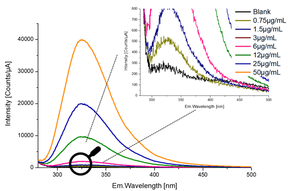
Figure 3. Fluorescence Emission spectra of transferrin at increasing concentrations (from 0.75 μg/ml to 50 μg/ml). Image Credit: HORIBA Scientific
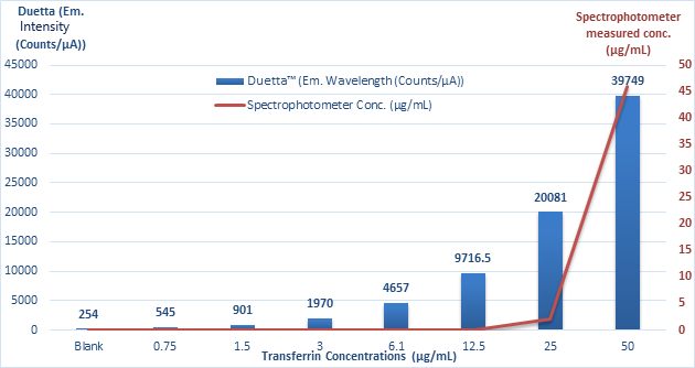
Figure 4. Comparison of Transferrin concentration measurements obtained with the spectrophotometer and Duetta instruments. Image Credit: HORIBA Scientific
Methods at a glance
For readers weighing whether these findings may translate to their own settings, the underlying experimental choices are central to interpreting the results.
- Analyte range: 50 µg/mL to 0.75 µg/mL transferrin prepared in 10 mM PBS; protein‑free buffer as blank, modeling an idealized low‑interference matrix.
- Excitation and detection: excitation at 250 nm; emission collected from 265-550 nm; two‑second CCD integration; 10 nm slits, balancing sensitivity with acquisition speed.
- Performance profile: fluorescence detected down to ~0.75 µg/mL; absorbance detection down to ~25 µg/mL on the comparator spectrophotometer, defining the relative limits of detection in this study.
- Optimization levers reported: longer integration, wider slits, and emission binning to enhance limit of detection, at the expense of throughput if pushed to extremes.
These parameters reflect a research-style optimization rather than a fully automated clinical workflow, but they offer a realistic starting point for hospital and reference labs exploring similar configurations.
Where this is relevant for health systems
For health systems, technology gains only matter when they intersect with patient pathways, operational pressures, and governance priorities such as equity and access.
- Use cases
- Pre-analytic scarcity: pediatric, oncology, and critical care samples with tight volume constraints, where re-draws carry clinical risk or ethical concerns.
- Population screening programs: iron deficiency and anemia of chronic disease where low‑level detection reduces repeat draws and helps maintain participation in large-scale initiatives.
- Biobank and translational research: longitudinal protein tracking at low abundance without labeling, preserving finite archived material while enabling protocol harmonization across sites.
- Operational impacts
- Lower detection limits can reduce re‑runs, aiding turnaround time, instrument utilization, and staffing efficiency in already stretched clinical chemistry sections.
- Dual‑mode systems can unify fluorescence and absorbance in one platform, simplifying maintenance, training, and capital planning for integrated delivery networks.
- Intrinsic fluorescence avoids labels, but matrix effects (hemolysis, lipemia, bilirubin) still require verification with patient specimens and may vary across populations, necessitating local risk assessment.
For executives and procurement teams, these technical advantages translate into decisions about how aggressively to modernize core laboratory infrastructure and which facilities should serve as early adopters within a network.
Quality and regulatory guardrails for laboratories
New detection approaches must fit within established clinical laboratory oversight. Labs evaluating fluorescence for protein quantification should map validation plans to recognized parameters and governance frameworks before use on patient specimens, aligning with institutional risk management and patient safety committees.
- Core analytical studies (typical expectations)
- Accuracy, precision (repeatability/reproducibility), reportable range, limit of blank/limit of detection, linearity, and analytical specificity.
- Carryover checks, matrix interference studies, and stability (sample and reagent).
- Calibration strategy and traceability, including acceptance criteria for lot changes and software updates.
- Regulatory context in the United States
- Clinical laboratories operate under the federal Clinical Laboratory Improvement Amendments (CLIA) and applicable accreditation (e.g., CAP, Joint Commission), which set expectations for method verification, documentation, and ongoing quality control.
- When a laboratory designs and validates its own method, governance requires documented risk assessment, change control, and competency, with clear labeling of results as laboratory‑developed where required and with medical staff informed of any analytical differences versus legacy methods.
- Instrument change management
- Side‑by‑side comparison with the laboratory’s current method using patient samples across medical decision ranges, with pre-specified acceptance criteria agreed by clinical leadership.
- Periodic proficiency testing or alternative assessment with defined acceptance thresholds, ensuring longitudinal comparability and defensibility in audits or inspections.
- Policy updates for critical values, repeat criteria, and reflex testing to ensure consistency across platforms and sites, particularly in multi-hospital systems.
How fluorescence can complement current clinical assays
Most hospital laboratories quantify transferrin via automated immunoassays or derive transferrin saturation using serum iron and total iron‑binding capacity. Fluorescence offers a label‑free option to study protein conformation and low‑level concentration changes. For labs already managing iron panels at scale, a dual‑mode instrument can serve as a validation and R&D workhorse while preserving absorbance workflows for high‑throughput routine testing.
- Potential roles
- Method development and verification for specialized protein studies, including evaluation of new biomarkers before they migrate onto high-throughput analyzers.
- Reflex testing design where low‑volume detection is necessary, such as second-line characterization in inconclusive or borderline iron studies.
- Education and training for cross‑functional teams in chemistry and molecular labs, building internal fluency with fluorescence-based techniques ahead of broader adoption.
For institutional decision-makers, this positions fluorescence not as a replacement for current immunoassays, but as an enabling capability that can shorten the distance between discovery work and clinically robust assays.
Important limitations to keep in view
- The comparison was performed with transferrin in buffer, not in human serum; real‑world performance can shift due to endogenous interferences, sample handling variability, and co‑morbid conditions.
- Detection limits alone do not define clinical utility; decision limits, imprecision at medical cut points, and commutability with existing methods are essential for safe interpretation and for maintaining longitudinal consistency in patient records.
- Throughput, ergonomics, service coverage, cybersecurity controls, and total cost of ownership remain material purchasing considerations for health systems, particularly where budgets and workforce capacity are under sustained pressure.
These constraints argue for phased adoption, with early implementation in reference or academic centers that can generate real‑world evidence before fluorescence-based workflows scale to community hospitals.
Instrument note for readers
The dual‑mode platform described is marketed as the Duetta spectrofluorometer, which combines simultaneous fluorescence and absorbance measurement in a single instrument. Fluorescence exploits intrinsic signals from tryptophan, tyrosine, and phenylalanine, enabling label‑free protein characterization at low concentrations. For health-system leaders, the key strategic question is not only whether an instrument like Duetta can see lower than current platforms, but how that capability will be governed, validated, and integrated into existing diagnostic pathways.
Related reading
